Lectures DCBT2023
Week 1:
Paul Verschure
Mehdi Khamassi
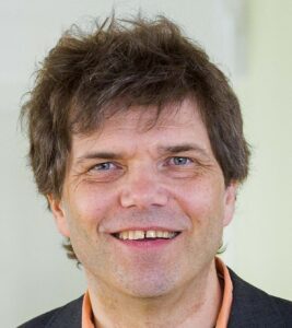
Fred Hamker
Floris de Lange
Tony Prescott
Wouter Serdijn
Sergi Bermúdez
Chris de Zeeuw
Ulyses Bernadet
Week 2:
Tatiana Korotkova
Michele Farisco
Florian Mormann
Chris Lewis
Heinz Beck
Vadym Gnatkovsky
Gaute Einevoll
Rick Helmich
Nick Ramsey
Michael Wenzel
Patrick Ruther
Goal-directed and habitual behavior
Fred Hamker 
I will initially introduce present state of the art in goal directed behaviour, as well as model-based on model free learning. Based on brain anatomy, I will derive a novel approach of goal directed behaviour and its learning. In short, I suggest that goal selection is implemented as part of the limbic loop connecting the hippocampus to the ventral basal ganglia, which reads-out of the virtualization in the memory system by the selection system to trigger the execution of a plan. Plan execution is then performed by further, hierarchically organised, cortex – basal ganglia loops, where lower level loops are constrained by objectives determined on higher level loops. Examples of simulation data from models are compared to experimental data.
—
Fred Hamker graduated in electrical engineering from the University of Paderborn in 1994 and received his PhD from the Faculty of Computer Science at the Technical University of Ilmenau in 1999. He worked at the JW Goethe University Frankfurt am Main and the California Institute of Technology (Pasadena, USA). In 2003, he founded a research group at the University of Münster in the Department of Psychology. Since 2009, he has held the professorship of Artificial Intelligence at the TU Chemnitz. Since 2010 he is associated with the Bernstein Center for Computational Neuroscience Berlin.
His research group takes a model-driven approach to studying how the brain works, with the goal of developing novel intelligent cognitive systems. Models are developed that help interpret psychological and neuroscientific data, thus contributing directly to the understanding of the brain. Likewise, research is being done on the transfer of neuroscientific knowledge into new concepts of artificial (brain-inspired) intelligence.
Model-based and model-free reinforcement learning mechanisms in brains and robot
Mehdi Khamassi
The reinforcement learning (RL) theory constitutes a framework for an artificial agent to learn actions that maximize rewards in the environment. It has been successfully applied to Neuroscience to account for animal neural and behavioral processes in simple laboratory tasks, such as Pavlovian and instrumental conditioning, and single-step economic decision-making tasks. It moreover became very popular due to its account for dopamine reward prediction error signals. However, more complex multi-step tasks, such as navigation and social interaction tasks, illustrate their computational limitations.In parallel, researches in engineering, in robotics in particular, have emphasized the complementarity between different learning strategies when facing complex tasks, and explored solutions to combine these different strategies. One central distinction is between model-based and model-free reinforcement learning strategies: In the former case, an agent learns a statistical model of the effects of its actions in the environment, and then use this model to plan sequences of actions towards desired goals. In contrast, model-free strategies are relevant when the environment statistics are too noisy to learn a good internal model. In this case, RL agents can rather learn local action values and adapt reactively in each state of the environment.In this presentation, I will show a series of work where we used a coordination of model-based and model-free reinforcement learning to account for a diversity of behavioral and neural observations in humans, non-human primates and rodents in different paradigms: Navigation, instrumental and Pavlovian conditioning. I will moreover present recent robotics results where the same algorithm with the same parameters produces optimal performance in simple navigation and social interaction tasks, with a drastically reduced computational cost compared to classical methods. Finally, I will show how the patterns of mental simulation within such internal models can mimic experimentally observed reactivations of the rodent hippocampus in spatial cognition tasks, and raise new predictions for future experiments.
—
Mehdi Khamassi is a research director employed by the Centre National de la Recherche Scientifique (CNRS), and working at the Institute of Intelligent Systems and Robotics (ISIR), on the campus of Sorbonne Université, Paris, France. He has a double background in Computer Science (Engineering diploma in 2003 from Ecole Nationale Supérieure d’Informatique pour l’Industrie et l’Entreprise, Evry with specialization in Artificial Intelligence and Statistical Modeling) and Cognitive Sciences (Cogmaster in 2003 from Université Pierre et Marie Curie (UPMC), Paris). Then he pursued a PhD in Cognitive Neuroscience at UPMC and Collège de France between 2003 and 2007. He serves as co-director of studies for the CogMaster program at Ecole Normale Supérieure (PSL) / EHESS / University of Paris Cité. He is editor-in-chief for Intellectica and serves as editor for several other journals, incl. Frontiers in Neurorobotics, Frontiers in Decision Neuroscience, ReScience X, and Neurons, Behavior, Data analysis and Theory. His main topics of research include decision-making and reinforcement learning in robots and humans, the role of social and non-social rewards in learning, and ethical questions raised by machine autonomous decision-making. His main methods are computational modeling, design of new neuroscience experiments to test model predictions, analysis of experimental data, design of AI algorithms for robots, and behavioral experimentation with humans, non-human animals and robots.
Rick Helmich 
Rick Helmich’s research delves into the intricate brain mechanisms underlying neurological movement disorders, with a primary focus on Parkinson’s disease and an emphasis on tremors. He explores the functional anatomy of neurological symptoms within the context of brain networks, utilizing an array of research tools such as functional MRI, EMG, SPECT, and TMS. His investigations encompass resting-state functional connectivity, effective connectivity through DCM, and task-based cerebral activity. The ultimate objective of his work is to unveil the origins of neurological symptoms, paving the way for innovative diagnostics and treatments for individuals grappling with movement disorders. Dr. Helmich’s impactful research has yielded valuable insights, as evidenced by his contributions to numerous publications, including studies examining stress and mindfulness in Parkinson’s disease and the amplification of Parkinson’s tremor under cognitive load.
Sergi Bermúdez
This seminar will explore the potential applications of virtual reality (VR) technology in stroke rehabilitation and fitness training. The integration of entertainment technology has been instrumental in advancing Virtual Rehabilitation research, leading to a surge in studies in this domain. The presentation will delve into the role of neurosciences in stroke rehabilitation research, the design methodology for rehabilitation applications, and the challenges of personalizing and customizing rehabilitation technology. The synergy between technology and neuroscience research is driving the field’s expansion. Developing VR applications involves formulating hypotheses based on technology’s underlying science and addressing customization to cater to each patient’s specific deficits, engagement, and comparison to conventional treatments in randomized control trials. This seminar will comprehensively review the approaches used in the last decade at the NeuroRehabLab for developing VR systems, encompassing hardware, software, design, and implementation methodologies.
—
Sergi Bermudez is an Associate Professor and researcher with expertise in Informatics, Human-Computer Interaction, Entertainment Technologies, and Neurorehabilitation. He holds key positions at the University of Madeira (UMa) as the coordinator of the NOVA LINCS FCT Center Branch and scientific director of LifeTech – the Quality of Life Technologies Center of Madeira. Sergi’s educational journey includes a degree in Telecommunications Engineering from UPC and a PhD from ETHZ. Throughout his career, he has conducted research at esteemed institutes in Europe and the USA, resulting in numerous publications and awards. Currently, he leads the NeuroRehabLab, which is dedicated to developing and evaluating innovative technologies and interventions to support the recovery and rehabilitation of individuals with neurological disorders and impairment
Cerebellar Orchestration of Motor Behaviour and Cognition
Chris de Zeeuw 
Over the past several decades, theories about cerebellar learning have evolved. A relatively simple view that highlighted the contribution of one major form of heterosynaptic plasticity to cerebellar motor learning has given way to a plethora of perspectives that suggest that many different forms of synaptic and non-synaptic plasticity, acting at various sites, can control multiple types of learning behaviour including cognitive processes. The challenge is to reconcile these different mechanisms and unite them into a single overall concept. In this lecture I review our current understanding of the changes in the activity of cerebellar Purkinje cells in different ‘microzones’ during various forms of learning. During the initial stages of relatively simple forms of learning, such as modification of sensorimotor reflexes, the simple spike activity of each microzone is bound to either increase or decrease. Instead, during more complex forms of learning, including planning and decision making, mixed sequences of increases and decreases of activity can occur within individual microzones, operating as ensembles in synergy. In either case, the upbound and downbound changes in activity in the microzones appear to depend on the directional and temporal demands of the downstream circuitry involved, continuously finetuning and orchestrating the optimal behaviour. In my lecture for the Donders Cognition, Brain and Technology Summer School I will highlight these fundamental principles as well as the clinical implications.
—
Dr. Christian de Zeeuw began his academic journey with a PhD in medicine Cum Laude under Jan Voogd at the University of Rotterdam, generating a new technology in which he demonstrated simultaneously the connectivity of nerve fibres and the identity of the neurotransmitters. He pursued neurophysiology under Rodolfo Llinas, where he unraveled the circuitry of the vestibulocerebellum underlying eye movement control and showed that this part of the brain generates predictions required for motor learning. In addition, he discovered the dendritic lamellar body. Transitioning to molecular biology with Frank Grosveld, he identified genes like CYLN2 linked to Williams Syndrome. Founding the Department of Neuroscience in 1998, which earned numerous awards, grants, and fellowships. Noteworthy accolades include the Beatrix Award, ERC-Adv grant, and Royal Dutch Academy of Arts & Sciences membership. He co-directed the Netherlands Institute for Neuroscience, initiated a cerebellar cognition group, and pioneered transformative programs. With commercial ventures like Neurasmus BV and BlinkLab, his numerous papers and citations delved into diverse neuroscience disciplines. De Zeeuw strives to unravel cerebellar disease mechanisms for innovative interventions, securing ExpertScape’s top global cerebellum researcher ranking.
Form and Function of Computer-generated Artificial Humans
Ulyses Bernardet
Ulysses Bernardet is a lecturer at Aston University in Birmingham, UK. Ulysses holds an MSc in psychology from the University of Zurich and did his doctorate in psychology while working at the Institute of Neuroinformatics, University of Zurich/Swiss Federal Institute of Technology in Zurich.
After his PhD, Ulysses was a senior researcher at the laboratory for Synthetic, Perceptive, Emotive, and Cognitive Systems, Universitat Pompeu Fabra, Spain. Until 2017 Ulysses was a postdoctoral researcher at the Simon Fraser University and a sessional lecturer at the University of British Columbia in Vancouver.
Ulysses has worked in the domain of motivation and behaviour regulation since his master’s in social motivation. During his PhD, he moved towards neuroscience and developed the large-scale neuronal systems simulator iqr. Using this tool, he did modelling and experimental work with robots on the path integration and behaviour regulation system in insects. As a postdoctoral researcher at the Pompeu Fabra University, he conceptualized and constructed the “eXperience Induction Machine” (XIM). Using XIM, Ulysses designed and conducted experimental studies in the domain of Humand-Computer-Interaction and Mixed-Reality. During his time in Canada, Ulysses’ focus shifted toward the development of realistic, autonomous virtual humans.
In his Cybernetic Human Lab at Aston University (cs.aston.ac.uk/chl) he researches virtual humans on the one hand for the application in computer games, training, etc., and on the other hand as a means to develop and test models of human cognition, affect, personality, and behaviour regulation.
Ulysses’ scientific approach is understanding humans by building them. For this purpose, his research practice brings together the domains of psychology and neuroscience with mixed and virtual reality.
Neil Levy
Eduardo Fernandez 
Professor of Cellular Biology, Chairman of the Department of Histology and Anatomy in the University Miguel Hernández (Spain) and Director of the Neuroengineering and Neuroprosthesis Unit at the Bioengineering Institute. He received a M.D. degree from the University of Alicante (1986) and a Ph.D. in Neuroscience with honors in 1990. He has been visiting professor at the University of Utah (USA), University of Oldenburg (Germany), Beth Israel Medical Deaconess Center (USA) and University of Vienna (Austria). His research interests is in developing solutions to the problems raised by interfacing the human nervous system and on this basis develop a two-way direct communication with neurons and ensembles of neurons. He is actively working on the development of neuroprostheses and brain-machine interfaces. In the last 5 years he has been using histological and electrophysiological techniques to asses the response to implantation and general biocompatibility issues regarding intracortical microelectrodes. He is also working on brain plasticity and reorganization in severe vision loss and developing non-invasive methodologies for the selection of appropriate candidates for implantation of visual neuroprosthesis
The synthetic psychology of theself: understanding human motivation and awareness through robotics
Tony Prescott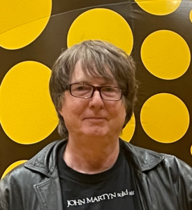
—
Tony Prescott (him/his) is a Professor of Robotics at the University of Sheffield who develops robots that resemble animals including humans. His goal is both to advance the understanding of biological life and to create useful new technologies such as assistive and educational robots. With his collaborators he has developed several animal-like robots including the whiskered robots Scratchbot and Shrewbot, and the pet-like robot MiRo-e which is currently in use for research and education and is being explored as potential therapeutic tool for children with anxiety. He has published over 200 refereed articles and journal papers at the intersection of psychology, brain science and robotics
Frits Matthijssen 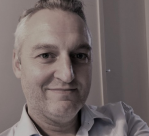
Frits Mattijssen is currently serving as a Knowledge Transfer Officer at Radboud University since March 2023. Before joining Radboud University, Frits Mattijssen spent almost 5 years at Bayer, where he held the position of National Relations Manager in Hoofddorp, Noord-Holland, Nederland. Prior to his corporate role, Frits served as a Medical Science Liaison for 2 years in Mijdrecht, Utrecht, Nederland. In this capacity, he engaged with healthcare professionals, contributing his scientific knowledge to support the development and promotion of cutting-edge medical solutions. Frits Mattijssen’s was a Postdoctoral Researcher at Helmholtz Zentrum München. Before that, he was a Postdoctoral Researcher at the DKFZ German Cancer Research Center. Frits Mattijssen’s academic journey began at Wageningen University & Research, where he earned his PhD in 2014. His doctoral research, titled “Functional characterization of the PPAR targets ANGPTL4 and HILPDA in lipid metabolism”.
Visual perception and consciousness – and new approaches for restoration when the eyes fail
Pieter Roelfsema 
A long-standing dream of scientists is to be able to directly project images from the outside world onto the visual brain, bypassing the eyes. This method could provide a solution for blind and visually impaired patients. It is the only possible solution for patients in whom the connection between eye and brain is lost so that a prosthesis in the eye is not an option.
I will first give an overview of the functioning of the visual cortex, which has low level areas for the analysis of simple visual features and higher areas for the analysis for more complex properties such as object category and face recognition. I will then discuss the mechanisms that determine whether a visual stimulus will reach consciousness or not. It is well established that the electrical stimulation of electrodes in the visual brain leads to artificial percepts called “phosphenes”. This method also works in patients who have been blind for decades. The goal of our own research is to bring a prosthesis for the visual brain closer. We implanted 1000 electrodes in the visual cortex to generate complex visual patterns. We demonstrated that this stimulation leads to interpretable images, in the same way that pixels form recognizable patterns on a screen. These new neurotechnological developments take important steps in the direction of prostheses that can restore a rudimantary form of vision.
—
Pieter R. Roelfsema received his MD degree in 1991 and his PhD degree in 1995. He moved to the Netherlands Institute for Neuroscience in Amsterdam in 2002 and became director in 2007. He is professor at the Free University of Amsterdam and at the AUMC in Amsterdam. He received a NWO-VICI award (2008) and two ERC-Advanced grants (2014 and 2022). Roelfsema studies visual perception, plasticity, memory and consciousness in the visual system of experimental animals, humans, and with neural networks. His main question is how neurons in different brain areas work together during seeing and thinking. Roelfsema studies how networks of neurons work together to perceive and solve cognitive tasks and how they configure themselves during learning. He develops the neurotechnology for high-bandwidth visual prostheses for blind people, aiming to restore a rudimentary form of sight. Roelfsema coordinates the Dutch neurotechnology initiative NeuroTech-NL. In 2019 he co-founded the start-up company Phosphoenix that aims to develop a visual brain prosthesis.
Michael Wenzel
Gaute Einevoll
Measurements of electric potentials from neural activity have played a key role in neuroscience for almost a century. Simulations of neural activity is an important tool for understanding such measurements and to make a quantitative link between what the neural network is doing and what is measured. Volume conductor (VC) theory is used to compute extracellular electric potentials such as extracellular spikes, MUA, LFP, ECoG and EEG surrounding neurons, and also magnetic signals such as MEG. In the lecture, the foundations of VC theory is outlined. Furthermore, examples are provided of how the theory is applied to compute spikes, LFP- EEG- and MEG signals generated by neurons and neuronal populations.
—
Gaute Einevoll is a professor of physics at the Norwegian University of Life Sciences and the University of Oslo working on brain physics, in particular, physics-type modelling of nerve cells, networks of nerve cells, brain tissue and brain signals. He is participating in the large-scale EU Human Brain Project started in 2013 and scheduled to end in 2023
Patrick Ruther
State-of-the-art stereo-electroencephalography (SEEG) probes are realized using precision machining of cylindrical electrodes lined up to electrode arrays of up to 18 recording and stimulation sites. These probes enable the integration of only a few micro-contacts to record single unit activity. The lecture presents different approaches to realize depth probes using micro-electromechanical system (MEMS) technologies based on flexible polymeric substrates. These technologies allow on the one hand cylindrical SEEG probes with up to 128 channels distributed along a cylinder with a diameter of 800 µm or highly flexible probes with unprecedented lengths of up to 35 cm targeting deep brain areas. The lecture will discuss the applied technologies and their respective limitations as well as requests for future systems optimizations.
—
Patrick Ruther received the Diploma degree in physics and the Ph.D. degree in mechanical engineering in 1993 and 1996, respectively. From 1996 to 1998, he held a post-doctoral position at the Research Center Karlsruhe, Germany, where he was developing LIGA-based microoptical components. Since 1998, he has been a Senior Scientist at the Department of Microsystems Engineering (IMTEK), University of Freiburg. His focus is on the design, fabrication, and characterization of CMOS-compatible MEMS devices, including microoptical functionality for biomedical applications, such as neuroscience and optogenetics. He coordinated the EU project NeuroSeeker targeting the next generation of neural probes comprising electronic and optical functionality. He is a Co- Founder of the spin-off company ATLAS Neuroengineering, Belgium. He is a member of the Cluster of Excellence BrainLinks-BrainTools at the University of Freiburg.
Vasso Giagka
Combined electrical and optical recording for long-term interrogation of multi-scale neural circuit dynamics
Chris Lewis 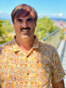
Adaptive behavior is enabled by the dynamic coordination of diverse signals across spatial and temporal scales. Integrating multi-channel electrical recordings with optical techniques can reveal dynamics invisible to standard, single-modality approaches. Fast dynamics recorded electrically can be related to simultaneously acquired optical signals from specified populations. We have developed flexible multi-electrode arrays that are chronically stable, permitting months long measurements of single units and local field potentials in the cortex, hippocampus, and thalamus of awake, behaving mice. We have combined chronic, multi-site electrophysiology with optical methods such as wide-field or 2-photon imaging, and fiber photometry cortex. We find diverse state-dependent patterns of coupling between concurrently acquired hippocampal and thalamic spiking activity and calcium dynamics in local circuits and across dorsal cortex. The combined application of electrical and optical methodologies provides a powerful tool with which to monitor and perturb brain-wide activity patterns. We believe the refinement of combined approaches will continue to reveal previously inaccessible and under-appreciated aspects of coordinated dynamics in the brain.
—
Christopher Lewis earned a BSc in Electrical Engineering and Computer Science with a second degree in Philosophy, Neuroscience and Psychology from Washington University, USA. During his studies he worked on vision and guidance systems for mobile robots in the Media and Machines group of Professor Robert Pless. Before pursuing graduate studies, he worked for some years as an engineer and software consultant in clinical and industrial settings. During his doctoral studies, he worked with a wide range of techniques and model organisms, including fMRI of clinical and non-clinical human cohorts, as well as large-scale electrophysiology and optogenetics in primates, cats, and rodents to investigate the effects of intrinsic brain activity on sensory coding and learning. He received his PhD in Neuroscience from Radboud University based on work performed at the Institute for Advanced Biomedical Technology in Chieti, Italy and the Ernst Strungmann Institute in Frankfurt, Germany. He is currently a staff scientist in the Helmchen lab at the Brain Research Institute, University of Zurich where he works with a small group developing novel electrode arrays to combine multi-channel electrophysiology with diverse optical methods. He is interested in understanding the brain-wide dynamics underlying perception and learning.
Artificial Awareness: moving from theoretical possibility towards empirical actuality
Michele Farisco 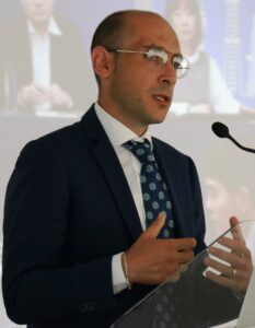
To make progress in the discussions about the theoretical possibility, logical coherence, conceptual plausibility, technical feasibility and empirical realism of artificial awareness, it is necessary to start from a preliminary theoretical reflection, which includes the clarification of the relevant terms as well as of the logical, theoretical and empirical conditions for artificial awareness to arise. In this talk I first sketch a conceptual analysis of key terms from consciousness research (including how the concept of awareness relates to the concept of consciousness) to then focus on the multidimensionality of consciousness and on the concept of consciousness profiles, introducing a relevant taxonomy. Against this background, I analyse the theoretical possibility of artificial awareness and reflect about how to identify (and possibly quantify) the dimensions composing a particular profile, arguing for the need of developing empirical, notably ecological indicators of consciousness in the case of artificial systems.
—
Human Brain Project and the EIC Pathfinder CAVAA project. He holds a MA in Philosophy from University of Naples “L’Orientale” in 2003, a PhD in “Ethics and Anthropology. History and Foundation” from University of Lecce in 2008, a Master degree in Biolaw from the University of Rome “Lumsa” in 2009, and a PhD in Neuroscience and Philosophy from Uppsala University in 2019. He spent time on an exchange grant from the European Neuroscience and Society Network within the European Science Foundation joining the Coma Science Group of the University of Liège (Belgium). He is the head of the “Science and society” research unit of Biogem Genetic Research Centre in Ariano Irpino (Italy). He is the author of four books and several articles about posthuman philosophy, philosophical, ethical and legal implications (ELSI) of genetics and neuroscience, consciousness (with a particular focus on disorders of consciousness), addiction, Artificial Intelligence, and neuroethics. He was awareded the title of Associate Professor of Moral Philosophy in Italy in 2015.
His current research focuses on consciousness, Artificial Intelligence, and reciprocal connection. Specifically, in collaboration with empirical scientists from inside and outside the HBP and the CAVAA project, his research aims at developing a philosophical and ethical framework for the experimental and computational explorations of cognition and consciousness. To illustrate, he is collaborating in the elaboration of concrete empirical, theoretical, and behavioural criteria for ascribing consciousness to people with disorders of consciousness, animals, and machines, and he is engaged in exploring the arising ethical issues.
Anna Kuppuswamy 
This lecture will present an overview of the field of post-stroke fatigue with an emphasis on investigations aimed at understanding the mechanisms of post-stroke fatigue. The lecture will be divided into 4 sections. In the first section, I will define the problem and briefly introduce methods to measure post-stroke fatigue. In the second section I will present the sensory attenuation model of fatigue and other mechanistic theories of post-stroke fatigue. The third section will deal with interventions and how best to tackle post-stroke fatigue. The fourth section will elaborate on unanswered questions, future directions and how understanding fatigue phenotypes is essential to progress our understanding of fatigue.
—
Anna Kuppuswamy is a Senior Lecturer at University of Leeds and an Honorary Principal Research Fellow at the Institute of Neurology. Her lab’s focus is on understanding the neural basis of fatigue in neurological disorders. Using multi-modal methodology ranging from neurostimulation, neuroimaging and behavioural methods, her lab probes the role of attentional, sensorimotor and limbic circuits in the experience of post-stroke fatigue.
Biofeedback-based audiovisual art generation
Héctor Lopez Carral
The aim of this project is project is to capitalize on closed-loop systems to augment artistic expression by giving a stronger emphasis to the performer’s subconscious activity. To achieve this, in this project we will use physiological sensors to obtain different signals (e.g., EEG, heart rate, electrodermal activity) and analyze them online to extract meaningful metrics about the user’s psychophysiological state. Then, we will use these metrics in real-time to adapt different parameters of a generative audiovisual artwork. The participants undertaking this project should have experience or interest in physiological signal processing and audiovisual arts (ideally, generative). Although the focus of this project is artistic, the technologies and methods employed can also be employed in other domains involving systems that dynamically adapt to their users.
Advancing Epilepsy Surgery: Integrating Stereoelectroencephalography (SEEG) for presurgical evaluation and quantitative analysis
Vadym Gnatkovsky
SEEG, a minimally invasive procedure involving the placement of depth electrodes within the brain, offers a platform for long-term monitoring and recording of epileptic activity. This provides crucial insights into the epileptogenic zone, facilitating accurate localization and characterization of the brain regions responsible for seizures. By accurately identifying the epileptogenic zone and preserving critical brain functions, patients experience improved outcomes and a reduced risk of postoperative deficits. The quantitative aspect of SEEG analysis adds a layer of sophistication to the evaluation process. Various quantitative techniques, including spike detection, functional mapping, source localization, and network analysis, collectively contribute to a comprehensive understanding of the complex neural dynamics underlying epileptogenic network. This lecture will expound upon several of these quantitative approaches.
—
Vadym Gnatkovsky is a Senior Researcher at the Department of Epileptology, University Hospital Bonn, Germany.
He studied Medicine from 1992 to 1999 at the Odessa State Medical University, Ukraine. In 2002, Dr. Gnatkovsky received his PhD in Pathological Physiology followed by postdoctoral training under the supervision of Dr. Marco de Curtis and Dr. Giuliano Avanzini at the Milan Istituto Neurologico “C. Besta”. His scientific interest addressed basic mechanisms underlying epileptic seizure development in experimental and human epilepsy as well as the identification and quantification of SEEG biomarkers of the epileptogenic zone. Ongoing studies to discover a potential mechanisms of seizure generation and propagation with human intracranial SEEG recordings. Aim of this work is studying intrinsic mechanisms of seizure generation, development end termination. He has been a member of the AES/ILAE’s Translational Task Force.
Technology for Bioelectronic Medicine
Wouter Serdijn
To better understand the brain and better treat brain disorders, it needs to be neuromodulated through ‘brain-like’ waveforms. Moreover, these waveforms need to be applied in a smart way, based on feedback, and in a closed-loop fashion. This requires sensing technology, not only for reading the electro-chemical signaling of the brain itself but also of other physiological parameters. Additionally, this feedback needs to be self-learning so it can learn to recognize the (personal) brain activity and connectivity characteristics that characterize a symptom and the intensity of a symptom. It then selects an optimal stimulation design to normalize the symptom by increasing or decreasing connectivity to change the network structure.
In this DCBT Summer School lecture I will address the technological aspects of how these ‘bioelectronic medicines’ can do this, what they will look like, and which future developments are needed to make them a reality. In particular, we will look into the network organization of the brain, power-efficient brain-like neuromodulation, flexible implant technology, and new modalities to interact with our electro-chemical mainframe, so that we can truly feel better.
—
Wouter Serdijn is a professor of bioelectronics, the head of the Section Bioelectronics of Delft University of Technology and as Medical-Delta professor also affiliated with the Neuroscience Department of the Erasmus Medical Center in Rotterdam, the Netherlands. He is fascinated by the electrical activity of the human body and the potential of bioelectronic medicine, also known as electroceuticals. Within the Medical Delta and the Dutch consortium NeuroTech-NL, he researches and develops bioelectronics for neuroscientific research and the development of bioelectronic medicine
Decoding Higher Order Cognition from Intracranial EEG
Christian Herff 
The decoding of higher order cognition directly from recordings of neural activity in the brain could enable a new generation of prosthetic devices. Accurate information about memory processes, reward perception and attempted speech and motor activity will allow targeted interventions and next-level human-computer interaction. In this presentation, I will present work with neurological patients that have electrodes implanted deep into their brains for clinical procedures. By piggybacking on these clinical routines, we are able to record high-fidelity neural activity across a variety of brain areas and align them to cognitive tasks. Through the application of machine learning, we are able to decode higher-order cognition from these recordings and process the output in real-time.
—
Dr. Christian Herff is an assistant professor in the School for Mental Health and Neuroscience at Maastricht University where he leads the invasive BCI research line. His research interest lays in the application of machine learning technology to neurophysiological data for Brain-Computer Interfaces and neuroscience research. With a particular focus on the decoding of speech processes from intracranial data, he tries to improve the lives of severely paralyzed patients while simultaneously improving our understanding of complex higher order cognition. He emphasizes the ability to achieve interpretable results based on computational models. In particular, visualization of complex dynamic models, such as deep neural networks, is of interest to him.
Simple pleasures: regulation of feeding and social interactions by subcortical neuronal circuits
Tatiana Korotkova 
Animals have to coordinate and prioritize multiple, at times competing basic needs to ensure survival and reproduction. Nutritional needs, such as hunger and thirst, ought to be balanced and weighed against competing needs like mating and voluntary exercise.Despite a long- known crucial role of the lateral hypothalamus (LH) and the reward system in the regulation of innate behaviours, the neuronal mechanisms enabling state-dependent representation and orchestration of multiple innate drives are not yet understood. Using optogenetics, chemogenetics, deep-brain calcium imaging and electrophysiology in freely behaving mice, we investigated functions of neuronal populations in LH and other subcortical brain regions in feeding and social interaction. We identified neuronal populations and circuits, which act in a complementary manner to enable the flexible fulfillment of multiple essential needs
—
Prof. Tatiana Korotkova studied biology with a focus on human and animal physiology in Lomonosov Moscow State University. During her Ph.D. under the supervision of Dr. R.E. Brown and Prof. H.L. Haas in Düsseldorf she investigated actions of various hypothalamic neuropeptides on aminergic nuclei, particularly on dopaminergic system. As a postdoc in the lab of Prof H. Monyer in Heidelberg, and later in a collaboration with Prof. T. J. Jentsch in Berlin she studied mechanisms of hippocampal network oscillations in behaving transgenic mice. In 2012-2017 T. Korotkova was a junior group leader at the Leibniz Institute of Molecular Pharmacology (FMP)/Neurocure Cluster of Excellence in Berlin, in 2017-2019 – a research group leader at MPI for Metabolism Research. Since 2019 T.Korotkova is a Full Professor and Managing Director at the Institute of Physiology, University of Cologne. She was a holder of the DFG and Schering foundation research stipends, and was awarded the Human Frontier Science Program (HFSP) Grant as well as the Junior Brain Prize by Lundbeck Foundation. The group of Tatiana Korotkova investigates neuronal mechanisms of innate behaviors, including feeding, social interactions and voluntary locomotion, in health and disease. Her research is supported by the European Research Council and by the German Research Organization (DFG). She supervised 13 Ph.D. and 12 M.Sc. students, and co-authored 29 peer-reviewed original publications, 4 reviews as well as one book chapter.
Lectures held in DCBT2022
Week 1: Network Neuroscience: from synapses to large-scale networks
Hippocampal-entorhinal circuits for spatial memory
The hippocampus is important for spatial and episodic memory. Place cells – the principal cell of the hippocampus – represent information about an animal’s spatial location. Yet, during sleep and rest, place cells spontaneously recapitulate (‘replay’) past trajectories. Replay has been hypothesised to serve a variety of functions in memory. In my talk, I will describe recent work I carried out, which showed replay may support a dual function: underpinning both spatial planning as well as the consolidation of new memories. Namely, we found during rest periods, place and grid cells from the deep medial entorhinal cortex (dMEC, the principal cortical output region of the hippocampus) replayed coherently. Importantly, putative dMEC replay lagged place cell replay by ~11ms; suggesting the replay coordination may reflect consolidation. Moreover, in a separate study, we found replay occurring just before movement to or upon arrival at a reward site preferentially depicted locations and trajectories consistent with the animals’ current task demands; perhaps indicative of spatial planning. However, we also found replay could dynamically ‘switch’ between a planning and consolidation mode in relation to engagement with task demands, and we found planning-like replay predicted the accuracy of imminent spatial decision. Finally, I will discuss unpublished work showing hippocampal-dMEC synchronisation in the theta-band may underlie hippocampal-dMEC replay coordination and ongoing work where we employ an ontogenetic approach to elucidating the neural circuit mechanisms of spatial memory.
—–
Dr. Hauður Freyja Ólafsdóttir received her PhD in Neuroscience from University College London (UK) in 2014 under the supervision of Prof. Hugo Spiers. Her thesis focused on the role of hippocampal place cell activity during sleep and wakefulness in spatial navigation. During her postdoc, in Dr. Caswell Barry’s (UCL) lab, she studied the contribution of hippocampal-cortical communication during different behavioral states to memory consolidation and spatial planning. In 2018 she received a Donders Mohrmann fellowship to start her own lab at the Donders Institute for Brain, Cognition and Behaviour (Radboud University, the Netherlands). The primary research objectives of her lab are to elucidate the neural circuit mechanisms underlying episodic and spatial memory, in development and adulthood, using state-of-the-art electrophysiological and imaging techniques.
Introduction to phase-amplitude coupling
This lecture covers mathematical concepts of coupling measures to estimate functional relations between neuronal oscillations partly within but mostly across different frequencies.
1. In the first part, I will give a very basic and largely qualitative introduction of fundamental measures to estimate the coupling of phases of oscillations at a specified frequency, with the most popular measures being the phase locking value (PLV) and coherence. I will also discuss the problem of volume conduction, meaning that functional relations can also be an artifact of signal mixing in sensor space, and how this can be addressed using e.g. the imaginary part of coherence.
2. In the main part of this lecture, I will discuss cross-frequency coupling and here basically phase-amplitude coupling. Several rather intuitive measures of phase-amplitude will be introduced. The main focus will then be on the relations between these coupling measures and bicoherence. Essentially a measure of coupling between phases at three different frequencies, which is a (normalized) third order statistical moment in the Fourier domain and which is analogous to coherence being a normalized second order statistical moment. It will be argued that the study of such relations is helpful to understand and optimize parameter setting for phase-amplitude coupling.
—–
Dr. Guido Nolte studied physics and made his PhD in 1995 at the University of Oldenburg/Germany. Since 1995 he has worked on methods development for the analysis of MEG and EEG data, in particular on forward and inverse calculations and on the construction of coupling measures. After working in Berlin, Albuquerque, Bethesda and Berlin again, he is now head of the MEG lab of the department of neurophysiology and pathophysiology at the UKE in Hamburg.
Models of large-scale brain dynamics
Brain activity exhibits chaotic signals comparable with the ones observed in networks of delay-coupled oscillators. On one side, transient brain rhythms are detected in EEG/MEG signals. On the other, fMRI reveals slow and spatiotemporally organized signal fluctuations. Using mathematical models of coupled oscillators, I will show how the network system can engage in a critical regime where intermittent cluster synchronization can generate signals sharing qualitative and quantitative features of human brain activity. I will present phenomenological insights obtained from models of coupled oscillators in the brain’s Connectome structure. Insights into general integrative processes of brain function will be discussed.
—–
Brain simulations: from neural dynamics to cognitive functions
The use of mathematical and computational models to study the brain has experienced a sharp rise in recent years as it constitutes a topic of interest, not only for neuroscience but for related disciplines like complex systems, nonlinear physics, psychology, and artificial intelligence. While many models have traditionally focused on ‘microscopic’ neural circuits (up to tens of thousands of neurons), novel data on detailed brain connectomes for human and no-human animals is facilitating the development of computational models of large-scale brain networks. In this lecture, I will cover recent work on large-scale brain modeling, and how it is used to understand the dynamics of neural activity observed in vivo and the perceptual and cognitive functions associated with such dynamics. In the first part, I will show how brain models are able to disprove predictions from local or microscopic computational models whose solutions do not hold then the large-scale recurrent connectivity of the brain is taken into account. A paradigmatical example is the propagation of sensory signals, a process traditionally studied with simplistic feedforward networks and which becomes dynamically unstable when real brain connectomes are considered. Proper solutions to this problem involve a strong balance between excitation and inhibition in brain networks. In the second part, I will show how this model can be extended to incorporate naturalistic neural oscillations, in an example of multiscale brain modeling. By constraining each level with neuroanatomical data and testing its dynamics with electrophysiological data, the multiscale model is able to reproduce a wide range of experimental observations across different spatial and temporal dynamics, and uncovering the emergence of functional hierarchical structures. Finally, in the third part, I will cover brain models which, besides realistic neural dynamics, can account for cognitive brain functions. In particular, I will introduce a data-constrained large-scale brain model able to explain the emergence of working memory –a particular type of short-term memory process in neural circuits –as a phenomenon distributed across large brain regions, in agreement with recent experimental evidence. This constitutes a new computational framework in which a large-scale model not only describes brain activity but also incorporates brain operations and function.
—–
Dr. Jorge Mejias is an assistant professor and head of the Computational Neuroscience Lab at the Cognitive and Systems Neuroscience Group at the University of Amsterdam in the Netherlands. With a background in physics and mathematics, he obtained a PhD in computational neuroscience from the University of Granada (Spain) in 2009. He then worked as a postdoctoral researcher at the University of Ottawa (Canada), New York University (USA), and the East China Normal University/ NYU Shanghai (China) before joining the University of Amsterdam in 2017. His research is focused on the theoretical and computational study of data-constrained multi-scale brain networks during perception and cognition, including brain functions such as working memory, multisensory integration, and predictive coding, as well as brain disorders that impair such functions. He also serves as an external member of the Institute Carlos I for Theoretical and Computational Physics in Granada, as a faculty member at the European Institute of Theoretical Neuroscience in Paris, and (currently) as director at the Organization for Computational Neurosciences (OCNS).
Excitation/inhibition balance as multi-scale mechanism regulating brain function
The opposing forces of excitatory (E) and inhibitory (I) signaling fundamentally shape activity at many levels of neuronal organization. Heuristic arguments have favored a certain “E/I balance” to be important for normal brain function and “E/I imbalance” is thought to characterize many neurological and psychiatric disorders. The concept of E/I balance, however, is not uniquely defined at the mechanistic level because of many contributing factors, such as the size, number, and cellular distribution of synapses, the decay time of synaptic currents, and network topology. Here, we explain a definition of E/I balance that is inspired by computational modeling of critical brain dynamics. In this framework, E/I balance is an emergent network property—a functional state characterized by high spatio-temporal complexity emerging in neuronal networks balancing between order and disorder. The definition enables measuring an “E/I ratio” at the network level, and we show its utility for understanding basic principles of information processing in neuronal networks and for studying brain disorders in which E/I balance may be disrupted.
—–
Dr. Klaus Linkenkaer-Hansen is Associate Professor in the Department of Integrative Neurophysiology at Center for Neurogenomics and Cognitive Research (CNCR) at VU Amsterdam. He received his MSc in Physics-biophysics from the Niels Bohr Institute (Copenhagen University, Denmark) in 1998. He received his PhD from Helsinki University of Technology (Finland) in 2002. Following post-doctoral fellowships at the Netherlands Institute for Brain Research and CNCR, he became team leader of the Neuronal Oscillations and Cognition group in 2008. His research has been funded by 35+ awards from a number of European funding agencies, including the VENI scholarship from the Netherlands Organisation for Health Research and Development (NWO/ZonMW), the Physical Sciences (NWO/EW), the Social Sciences (NWO/MaGW), and the Technology Foundation (NWO/STW) of the Netherlands Organization for Scientific Research (NWO). His research is focused on the complex dynamics of neuronal oscillations and its implications for cognition in health and disease. Theories and methods from the physics of self-organization and complexity have guided his multi-disciplinary research since 1998. His research has shown that the complex temporal structure of ongoing oscillations is rich in information about the functional state and structure of the underlying neuronal networks.
Adaptive stimulation strategies
Stimulation-based therapies for brain disorders provide an exciting avenue due to their focal and reversible nature. Current approaches face limits of side-effects, and effectiveness being restricted to a small subset of the patient population. During this talk, I will highlight recent work on developing and testing new therapies that aim to overcome these challenges.
—–
Dr. Hayriye Cagnan studied Electrical and Electronics engineering at Cornell University and specialized in signal processing and biomedical engineering (2000-2004). In 2004, she was accepted to the M.Sc. programme in Engineering and Physical Science in Medicine at Imperial College. Hayriye completed her Ph.D. in Neuroscience in a joint placement between the University of Amsterdam and Philips Research Laboratories in 2010. Subsequently, she joined the University of Oxford and worked on tremor pathophysiology. In 2015, Hayriye was awarded an MRC Skills Development Fellowship in Biomedical Informatics and worked on theoretical modelling of disease circuits. Hayriye was awarded an MRC Career Development Award in 2018 to establish her research group on Dynamic Neuromodulation.
Entering the neurorehabilitation market
Going from the lab and controlled clinical trials to the real world poses additional challenges to neurorehabilitation solutions. In this talk, we will cover some of the main aspects neurorehabilitation products and companies must address for a successful commercial uptake.
—–
Eng. Santiago Brandi is an expert in technology for Neurorehabilitation and is the CEO of the spin-off company Eodyne.
Week 2. Brain Networks in Health and Disease: the Future of Neurotechnologies.
Past, presence, and future from neuroConn’s perspective.
The talk will focus on the history of neuroConn, the approach of neurocare for innovating mental health, and the future of having integrated solutions available for everyone.
—–
Dr. Klaus Schellhorn has long-standing experience in neurostimulation & neurofeedback technology. He is the managing director of the company neuroConn GmbH that he co-founded as a spin-off of the Biomedical Engineering Department at the Technical University of Ilmenau in 2000.
Computational models to study the Parkinsonian brain and the mechanisms of neurostimulation techniques
Surgical treatment of neurological disorders like Parkinson’s disease, dystonia, and epilepsy was, until recently, mainly based on applying lesions at specific parts of the brain. While these procedures nowadays have been replaced by more reversible neurostimulation methods, most therapies for brain disorders are still based on trial-and-error, and effective mechanisms remain unknown. Using computational modeling can help us provide insight into neuronal network processes and interactions underlying normal and abnormal behaviour, as well as the mechanisms of therapeutic methods. While the difficulty in modeling is determining how much complexity needs to be included to simulate the aspects we are interested in as realistically as possible, it allows us to easily test new stimulation paradigms and stimulation targets. However, validation is essential for computational models to become useful tools for research purposes and clinical applications. In this presentation, two types of computational modeling will be presented and discussed in relation to experimental or clinical data: 1) 3D volume conduction modeling to study the local stimulation effects of deep brain stimulation (DBS) and motor cortex stimulation (MCS) in Parkinson’s disease; 2) neuronal network modeling, at a microscopic (cellular) and macroscopic (system) level, to study the behaviour of (part of) the basal ganglia-thalamocortical network under normal and Parkinsonian conditions and the effect of DBS.
—–
Dr. Ciska Heida is an Associate Professor in the Biomedical Signals & Systems group at the University of Twente, The Netherlands. She has a background in Electrical Engineering and received her PhD in the field of neurotechnology at the University of Twente. Her current research interests focus on increasing our understanding of the central mechanisms of human motor control, the pathophysiology underlying movement disorders, and the development and application of neuromodulation techniques for restoring motor control. Her research methods consist of computational modeling of (part of) the brain networks involved in motor control, and experimental research including movement tests performed by patients while recording brain activity. Her teaching activities are related to signal analysis of biomedical/clinical data and bioelectromagnetics.
Neurotechnology for Parkinson’s Disease
Parkinson’s disease is one of the fastest-growing neurodegenerative diseases in the world. Advances in neurotechnology are necessary for a better understanding of the fundamental neurobiological mechanisms causing the disease, as well as for new approaches for diagnosis, and new devices for monitoring and treatment. In this seminar, I will provide an in-depth discussion of several of the new directions in this research field.
—–
Dr. Richard van Wezel is professor in Visual Neuroscience at the Radboud University Nijmegen and professor in Neurophysiology at Twente University (MedTech Center). He was director of the Donders Centre for Neuroscience in Nijmegen, and currently, he is vice-dean of Research at the Science faculty of Radboud University. Prof. van Wezel received many prestigious personal and team research grants (VIDI/NWO, High Potential UU, INTENS and other grants). He is co-chair of NeuroTech-NL (https://neurotech-nl.com/), a nation-wide consortium to promote Neurotechnology in the Netherlands. His main interest is in neurophysiology, visual perception, and brain plasticity and the applications of his fundamental research for clinical applications with a current applied focus on developing neurotechnology for Parkinson patients.
Feedforward and feedback processes that underlie visual perception
Visual perception is the result of an interaction between the bottom-up information being conveyed via the retina and top-down processes reflecting our prior knowledge and expectations. In this lecture, I will explore how feedforward signals, either natural or artificially induced, can lead to conscious perception and how feedback signals organize our perception into the rich 3D percept we enjoy every day.
—–
Dr. Matthew Self is a senior researcher at the Netherlands Institute for Neuroscience in Amsterdam. After completing his studies at Cambridge University, he joined Professor Semir Zeki’s lab at University College London to study the integration of different visual features using fMRI. He then joined the lab of Pieter Roelfsema in Amsterdam to carry out post-doctoral work on the processing of feedback signals in the different layers of the visual cortex. His current work focuses on how feedback and feedforward processes interact in the visual cortex to produce visual perception.
The cerebral network of Parkinson’s disease tremor: a talk about effective connectivity
Parkinson’s disease is characterized by the degeneration of dopaminergic cells in the midbrain, which leads to dopamine depletion in the striatum. One of the cardinal motor symptoms of Parkinson’s disease is a low-frequency (4-6 Hz) resting tremor. In the past, several studies have pointed toward the involvement of two cerebral circuits in the pathogenesis of Parkinson’s disease resting tremor: basal ganglia and a cerebello-thalamo-cortical circuit (consisting of cerebellum, thalamus and motor cortex). The exact circuit-level architecture of causal interactions between both circuits has long remained unclear, which has been a topic for our research over the last couple of years. In this presentation, we will talk about how we used functional MRI together with computational modelling (i.e. Dynamic Causal Modelling) to study the cerebral network of Parkinson’s disease resting tremor. We will focus on tremor-related cerebral activity and how different cerebral regions influence each other, i.e., how they are effectively connected. We propose the dimmer-switch hypothesis where the basal ganglia initiate a tremor episode, and the cerebello-thalamo-cortical circuit produces tremor amplitude. Next, we will discuss how this system-level framework has formed the basis to study several clinical characteristics of Parkinson’s disease tremor, such as the influence of dopaminergic medication and cognitive stress. Finally, we will discuss how this knowledge can be used to improve the therapy of Parkinson’s disease tremor.
—–
Dr. Michiel Dirkx is a post-doctoral researcher at the Donders Centre for Cognitive Neuroimaging and a neurologist in training at the Radboud University Medical Centre. He obtained his medical degree at the Radboud University Nijmegen in 2018, and his PhD thesis (“Neural mechanism of Parkinson’s tremor”) in 2020. Since 2020 he has worked as a post-doctoral researcher in the group of dr. Rick Helmich (“Systems Neurology”) where he focusses on the pathophysiology of movement disorders such as Parkinson’s disease. For this, he uses methods such functional MRI, electrophysiological tools such as EMG/accelerometry and computational modelling (Dynamic Causal Modelling).
Lessons for neurorobotics from bio-inspired learning and control principles of the central nervous system
This talk will present some principles for the design of neurorobotics systems to reproduce animal behaviours and test brain theories. I will for example refer to the embodied intelligence principle that refers to the capability of living systems to learn, act, and adapt during interactions with dynamic environments. Neuro-robots become able to learn and adapt their tasks in complex and changing environments by means of bio-inspired control systems in which the brain is embedded. We will discuss how computational models in motor control and model learning architectures, including the internal model learning theory, can be applied to build bio-inspired control systems. Results represent a step closer towards a better understanding of the underlying neural mechanisms for motor control and learning, but also the starting point for achieving collaborative robots acting in complex applications such as manufacturing, assistive living, or as a tool for discovering the root causes of neurodegenerative diseases.
—–
Dr. Silvia Tolu is an Associate Professor and currently leads the Neurorobotics team at the Technical University of Denmark (DTU). She holds a Ph.D. degree in Neuromorphic Computing from the University of Granada (Spain). Her main research experience is in bio-inspired control for robots, compliant human-robot interactions, computational models of the cerebellum, and machine learning. Her research focuses on developing brain-based methods and technologies for enabling autonomy in robots while they act in realistic, dynamic, and rich sensory environments. Her vision is to open revolutionary robotics paradigms and to discover novel methods for diagnosis and rehabilitation of neuro-degenerative diseases. In 2016, she was granted with an IF-EF H2020 Marie Sklodowska-Curie Fellowship (BIOMODULAR, Project ID: 70510 2017-2019). She has been principal investigator in the Co-Design Project 2 (CDP2 Human Brain Project HBP-SGA2) about cerebellar implementations in robotic systems (2018-2020). Silvia has published in influential journals and proceedings in both robotics and computational neuroscience and has served as reviewer for top-ranked conferences and journals. She is currently in the Editorial Board of Frontiers Neurorobotics.
Multimodal Neuroimaging with CURRY
This presentation will cover how the CURRY software integrates multiple imaging modalities (MRI, CT, PET, SPECT, fMRI, DTI) together with MEG/EEG data for offering various insightful possibilities when analyzing the brain. We will go through practical scenarios in which CURRY is useful for obtaining the most out of the available data: (a) reviewing multiple co-registered imaging modalities simultaneously, (b) creating anatomically accurate and subject-specific head models from MRI and DTI in order to perform source analysis, (c) pre-operative planning of stereo-EEG (intracranial) electrode implantation and post-operative evaluation, (d) combined MEG/EEG data and source analysis.
—–
Dr. Fernando Gasca graduated from the Iberoamerican University in Mexico City with a degree in Biomedical Engineering. He was co-founder of CODE Ingeniería, a Mexico City-based technology development company, where he worked as an engineer. He later received his Ph.D. in Neuroengineering from the University of Lübeck, Germany. His research focused on the modeling of transcranial stimulation techniques. Since 2014 he joined Compumedics Europe GmbH, Compumedics Neuroscan, as part of the CURRY development team in Hamburg, Germany. He has been involved in the development of the Image and Signal Processing modules, as well as the Finite Element Method (FEM) functionality and automatic spike and seizure detection. He is presently at
Brain networks in stroke and cerebral small vessel disease
I will discuss MRI connectome analysis and clinical implications in our prospective stroke cohorts and the Hamburg City Health Study (HCHS), the local, large-scale epidemiological study.
—–
Dr. Bastian Cheng is a consultant neurologist and associate professor at the University Medical Hospital in Hamburg. His clinical focus lies on the diagnosis and treatment of patients with stroke and cerebrovascular diseases. Scientific key interests are structural and functional magnetic resonance imaging in stroke, multimodal approaches of lesion-symptom inference as well as modelling and analysis of localized and large-scale structural brain networks in stroke and cerebrovascular disease relating to clinical recovery.